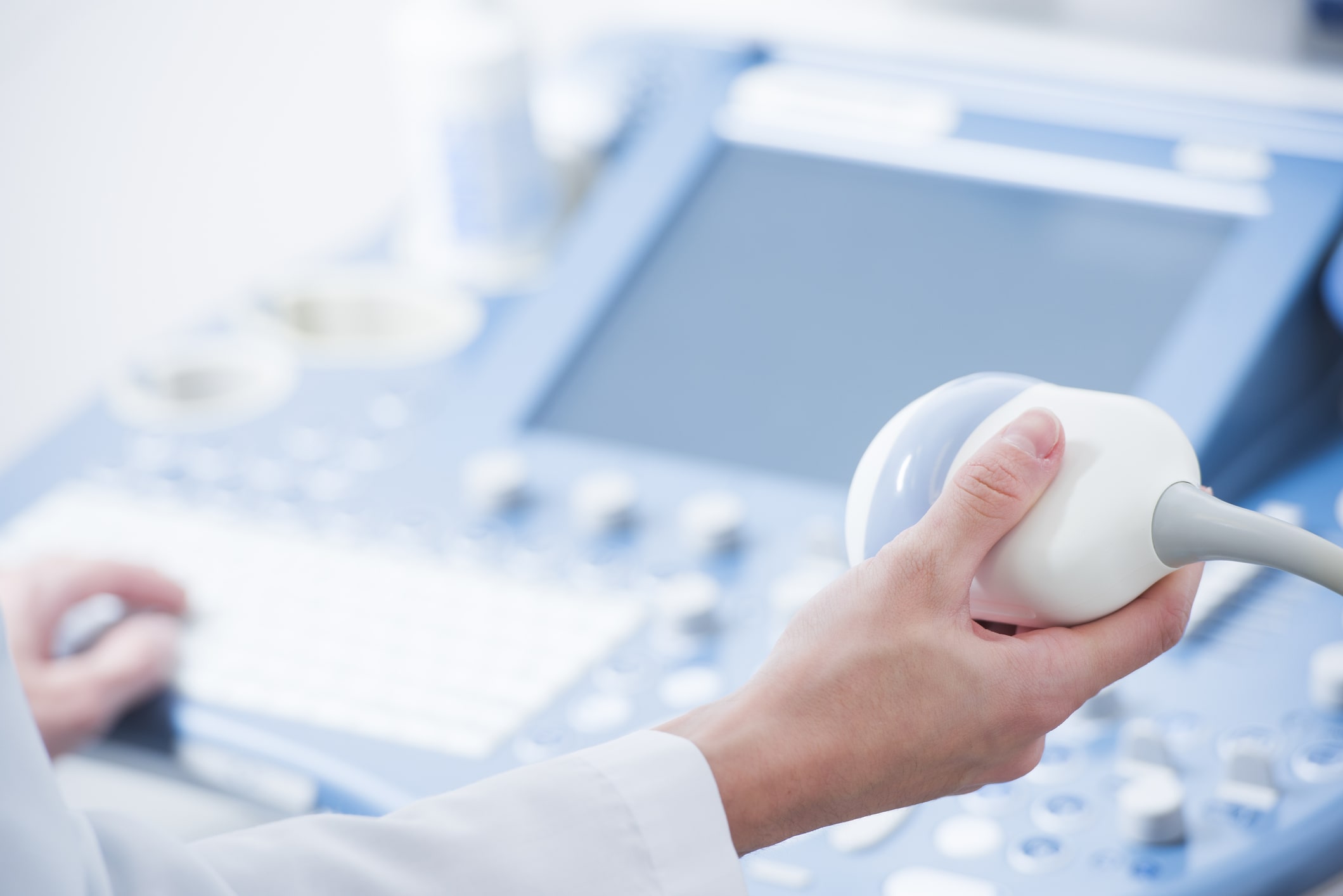
Picture of a transducer probe used during an ultrasound exam. High-frequency sound waves are reflected by tissues in the body.

Five Amazing Ultrasound Inventions Set To Change The World And Not A Pregnancy Scan In Sight
Rectangular at the top and curved at the bottom it is a phased array.
. What are two ways in which ultrasound technology produces images. Once the raw data are processed the CPU forms the image on the monitor. An ultrasound scan uses high-frequency sound waves to create images of the inside of the body.
Physics 26042021 2100 jocelynmarquillo1 What are two ways in which ultrasound technology produces images. Body tissues absorb high-frequency sound waves producing an image. If the shapes are different eg.
Ultrasound imaging uses sound waves to produce pictures of the inside of the body. Which two statements describe how ultrasound technology produces an image of part of the body. The strength amplitude of the.
It is the transducer that emits sound waves at frequencies ranging between 1 and 10 MHz megahertz. It works by transmitting sound waves into an object using a piezoelectric transducer then using the same transducer to pick up sound reflected from structures within the object. It then converts the reflected signals into audible sounds that the human brain can learn to process into a detailed mental image of the environment.
Linear arrays step the beam along the transducer and produce images with full image width up to the skins surface. By detecting the frequencies of emitted sound waves D. As a general rule if the shape at the top of the images matches the shape at the bottom of the image it is a sequential array.
By measuring the time it takes for reflected sound waves to be. Linear array also called sequential array phased array. The ultrasound image is produced based on the reflection of the waves off of the body structures.
Functional ultrasound combines information such as the movement and velocity of tissue or blood softness or hardness of tissue and other physical characteristics with anatomical images to create information maps. By using sound in frequencies that reflect off body tissues B. It helps diagnose the causes of pain swelling and infection in the bodys internal organs and to examine an unborn child fetus in pregnant women.
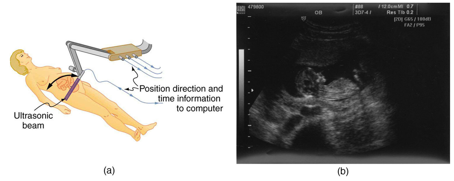
Display - displays the image from the ultrasound data processed by the CPU keyboardcursor - inputs data and takes measurements from the display disk storage device hard floppy CD - stores the acquired images printer - prints the image from the displayed data Transducer Probe The transducer probe is the main part of the ultrasound machine. In infants doctors commonly use ultrasound to evaluate the brain hips and spine. By using sound in frequencies that reflect off body tissues D.
Real-time ultrasound images are integrated images resulting from reflection of organ surfaces and scattering within heterogeneous tissues. By using sound in frequencies that reflect off body tissues B. By detecting the frequencies of emitted sound waves C.
By the absorption of sound waves in the body U D. Question 1 of 10 What are two ways in which ultrasound technology produces images. These waves dont return to the probe and are therefore wasted.
By the absorption of sound waves in the body O D. Ultrasound scanning is an interactive procedure involving the operator patient and ultrasound instruments. By measuring the time it takes for reflected sound waves to be detected Answers.
That is the body absorbs the ultrasound energy making the waves disappear. This can also include two-way conversations which can help to ensure that the images are being acquired correctly. An image is created based on the amount of time it takes for a sound wave to return.
The rapid development of ultrasound technology is expanding its global footprint beyond what could have. Answered Which two statements describe how ultrasound technology produces an image of a baby before it is born. By the absorption of sound waves in the body B.
Transducers can produce an ultrasound beam in two ways. When ultrasound enters the body some of it undergoes. Reflection Attenuation Some of the ultrasound waves are attenuated.
Magnetic Resonance Imaging is a non-invasive diagnostic technique that combines a large powerful magnetic field with radio frequencies to gaze into the human body without using x-rays. By using sound in frequencies that reflect off body tissues B. The resulting voltages in the transducer are then converted into electrical signals from which images can be reconstructed by a computer.
The first commercially available handheld B-mode scanner was produced in 1963 6. In time this technology could become more practical and portable perhaps even one day incorporated into specially designed glasses. The CPU can also store the processed data andor image on disk.
B-mode brightness-mode scanners are what is most often thought of when one refers to ultrasound. It is suitable for use during pregnancy. The type of emission imagining produces detailed pictures of organs soft tissues bone and other internal body structures without ionizing radiation.
By detecting the frequencies of emitted sound waves C. Ultrasound application allows for noninvasive visualization of tissue structures. B-mode produces a two-dimensional reconstructed image of internal body structures based on reflected sound waves.
Ultrasound scans or sonography are safe because they use. By detecting the frequencies of emitted sound waves O C. The transducer is passed over the area of the body that covers the internal structures to be imaged.
In some cases 3d and 4d ultrasound pictures may reveal abnormalities not readily seen using 2d ultrasound. By measuring the time it takes for reflected sound waves to be detected C. Anatomical ultrasound produces images of internal organs or other structures.
In turn the sound waves are reflected back to the transducer after they bounce off the structures that are the focus of the ultrasound. By the absorption of sound waves in the body Answers. Question 6 of 10 What are two ways in which ultrasound technology produces images.
Low-frequency sound waves are reflected by tissues in the body. By updating 3d ultrasound images in rapid succession sonographers can also create 4d ultrasound pictures. The transducer pulse controls allow the operator called the ultrasonographer to set and change the frequency and duration of the ultrasound pulses as well as the scan mode of the machine.
An image is created based on the frequency of reflected X-rays. By measuring the time it takes for reflected sound waves to be detected Answers Answer from. The amount of time it takes for a wave to return can be used to create an image.
In the 4d ultrasound the fourth dimension time adds movement and creates the most realistic representation of all.
What Can An Ultrasound Detect Envision Radiology
Ultrasound Physics

Pin On Health

Ultrasound Scans How Do They Work


0 Comments